


Cardiapoda
Roger R. Seapy

- Cardiapoda placenta (Lesson 1830)
- Cardiapoda richardi Vassière 1904
Introduction
The genus Cardiapoda is similar in general body shape to Carinaria, but differs most conspicuously in lacking a macroscopic shell that covers the visceral nucleus; instead the shell is microscopic (only slightly larger than the larval shell) and is imbedded in the dorsal portion of the digestive gland in the visceral nucleus. Both the right and left tentacles are well-developed and of equal size, unlike Carinaria (in which the right tentacle is greatly reduced or vestigial) and Pterosoma (where the right tentacle is absent). The cutis is thin, unlike the thick, gelatinous cutis in Carinaria and Pterosoma.
Brief Diagnosis
A carinariid genus with:
- Visceral nucleus exposed; not covered by a macroscopic shell
- Shell microscopic and imbedded in dorsal part of visceral nucleus
- Right and left tentacles well-developed and of equal size
- Tail ends either in a contractile fan-shaped expansion or a filamentous extension that can be greatly lengthened or shortened
Characteristics
- Body morphology
- Visceral nucleus not covered by shell, as in Carinaria and Pterosoma
- Gills either numerous, forming a crest on the visceral nucleus, or few, located at the entrance to the mantle cavity
- Visceral nucleus stalk directed posteriorly
- Right and left tentacles of equal size and well developedClick on an image to view larger version & data in a new window
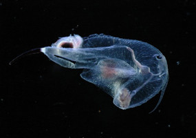
Figure. Frontal view of Cardiapoda richardi. © L. Madin
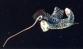
- Tail with a small dorsal crest that terminates either in a fan-shaped expansion that can be contracted or expanded (C. placenta) or a filamentous extension that can be greatly lengthened or shortened (C. richardi)Click on an image to view larger version & data in a new window
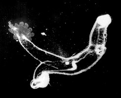
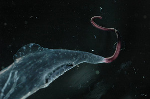
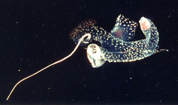
Figure. Lateral views of Cardiapoda placenta (left) with the terminal fan-shaped expansion and Cardiapoda richardi (right) with the low dorsal crest, elongate mid-ventral slit, and contracted reddish-brown filamentous tail extension. © R. Gilmer and © L. Madin, respectively.
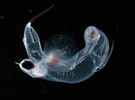
- Cutis thin; in contrast with the thick, gelatinous cutis of Carinaria and PterosomaClick on an image to view larger version & data in a new window
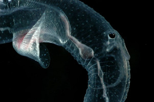
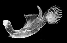
Figure. Swimming fin, head region and proboscis in Cardiapoda richardi. Note the folds in the thin cutis in the ventral part of the trunk and proboscis, resulting from ventral flexion of the proboscis. © L. Madin
- Visceral nucleus not covered by shell, as in Carinaria and Pterosoma
- Shell
- The adult shell is microscopic and consists of the protoconch (the larval shell following metamorphosis) and a rudimentary teleoconch. The teleoconch is produced at right angles to the shell aperture, forming an outwardly-radiating shelfClick on an image to view larger version & data in a new window
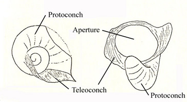
Figure. Drawings of the microscopic adult shell in Cardiapoda placenta, viewed from the right side and aperture (left and right, respectively). Shell diameter about 0.8 mm. Drawings modified from Tesch (1949) by addition of labels.
- Larval shell globular. Right side of shell with two spiral ridges on the second spire whorl. Left side of shell with umbilicus open and about 13 radiating ridges on umbilicus wall; aperture wide, slightly greater than 1/2 shell diameter Click on an image to view larger version & data in a new window
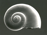
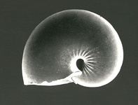
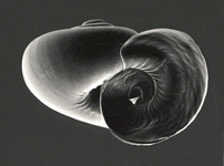
Figure. Scanning electron micrographs of the larval shell of Cardiapoda richardi, viewed from the right side, left side, and aperture. Shell diameter = 0.6 mm. © R. Seapy
- With growth of the animal, the microscopic shell becomes imbedded in the digestive gland, with only the top of the protoconch visible
- The adult shell is microscopic and consists of the protoconch (the larval shell following metamorphosis) and a rudimentary teleoconch. The teleoconch is produced at right angles to the shell aperture, forming an outwardly-radiating shelf
Comments
The genus Cardiapoda includes two species, which can be distinguished by the following characters:
| Species | Maximal body length | Number of gills | Location of gills | Fin sucker | Structures at end of tail | Ventral tail structure | Eye morphology | ||
| C. placenta | ca. 110 mm | >20 | form a crest on surface of visceral nucleus | present in both sexes | 12 finger-like expansions that can be contracted or expanded, forming a fan | absent | shape triangular, as in other carinariids | ||
| C. richardi | ca. 30 mm | 8 | at entrance to mantle cavity on visceral nucleus | present only in males | reddish-brown filamentous tail extension | flat membranous structure that can be extended from elongate ventral slit | shape oval, with lens resting in cup of black pigmented tissue | ||
References
Lalli, C. M. and R. W. Gilmer. 1989. Pelagic snails. The biology of holoplanktonic gastropod snails. Stanford University Press, Stanford. 259 pp.
Richter, G. and R. R. Seapy. 1999. Heteropoda, pp. 621-647. In: D. Boltovskoy (ed.), South Atlantic Zooplankton. Backhuys Publishers, Leiden.
Tesch, J. J. 1949. Heteropoda. Dana Report 34, 53 pp., 5 plates.
Title Illustrations

| Scientific Name | Cardiapoda placenta |
|---|---|
| Location | Florida Current |
| Specimen Condition | Live Specimen |
| Sex | Female |
| Life Cycle Stage | adult |
| View | right side |
| Size | 65 mm |
| Copyright | © Ronald Gilmer |
| Scientific Name | Cardiapoda richardi |
|---|---|
| Location | Florida Current |
| Specimen Condition | Live Specimen |
| Sex | Female |
| Life Cycle Stage | adult |
| View | left side |
| Size | 65 mm |
| Copyright | © Ronald Gilmer |
| Scientific Name | Cardiapoda richardi |
|---|---|
| Location | Sargasso Sea |
| Specimen Condition | Live Specimen |
| Sex | Female |
| Life Cycle Stage | adult |
| View | ventral |
| Copyright | © L. Madin |
About This Page

California State University, Fullerton, California, USA
Correspondence regarding this page should be directed to Roger R. Seapy at
Page copyright © 2008
 Page: Tree of Life
Cardiapoda .
Authored by
Roger R. Seapy.
The TEXT of this page is licensed under the
Creative Commons Attribution License - Version 3.0. Note that images and other media
featured on this page are each governed by their own license, and they may or may not be available
for reuse. Click on an image or a media link to access the media data window, which provides the
relevant licensing information. For the general terms and conditions of ToL material reuse and
redistribution, please see the Tree of Life Copyright
Policies.
Page: Tree of Life
Cardiapoda .
Authored by
Roger R. Seapy.
The TEXT of this page is licensed under the
Creative Commons Attribution License - Version 3.0. Note that images and other media
featured on this page are each governed by their own license, and they may or may not be available
for reuse. Click on an image or a media link to access the media data window, which provides the
relevant licensing information. For the general terms and conditions of ToL material reuse and
redistribution, please see the Tree of Life Copyright
Policies.
- First online 12 February 2008
- Content changed 12 February 2008
Citing this page:
Seapy, Roger R. 2008. Cardiapoda . Version 12 February 2008. http://tolweb.org/Cardiapoda/28742/2008.02.12 in The Tree of Life Web Project, http://tolweb.org/


