
Click on an image to view larger version & data in a new window
 Figure
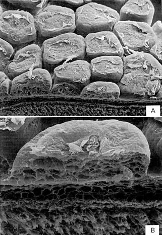
. Scanning electron micrographs of the dermal cushions of
Pholidoteuthis adami, 179
mm ML.
Top (A) - External appearance of pads. Field of view from left to right is 2.6 mm.
Bottom (B) - Cross-section of a cushion. The cushion is highly vacuolated as are the dermal cushions of
Lepidoteuthis grimaldii. Field of view is 0.7 mm. Photographs from Goldman, 1995.
References
Goldman, D. A. 1995. A juvenile of the scaled squid, Pholidoteuthis adami Voss, 1956 (Cephalopoda: Oegopsida), from the Florida Keys. Proc. Biol. Soc. Wash. 108: 136-146.
About This Page

University of Hawaii, Honolulu, HI, USA
National Museum of Natural History, Washington, D. C. , USA
Page copyright © 1999 and
 Page: Tree of Life
Pholidoteuthis adami Dermal Cushions
Authored by
Richard E. Young and Michael Vecchione.
The TEXT of this page is licensed under the
Creative Commons Attribution-NonCommercial License - Version 3.0. Note that images and other media
featured on this page are each governed by their own license, and they may or may not be available
for reuse. Click on an image or a media link to access the media data window, which provides the
relevant licensing information. For the general terms and conditions of ToL material reuse and
redistribution, please see the Tree of Life Copyright
Policies.
Page: Tree of Life
Pholidoteuthis adami Dermal Cushions
Authored by
Richard E. Young and Michael Vecchione.
The TEXT of this page is licensed under the
Creative Commons Attribution-NonCommercial License - Version 3.0. Note that images and other media
featured on this page are each governed by their own license, and they may or may not be available
for reuse. Click on an image or a media link to access the media data window, which provides the
relevant licensing information. For the general terms and conditions of ToL material reuse and
redistribution, please see the Tree of Life Copyright
Policies.


